Dengue Fever-Induced Complete Heart Block Requiring Permanent Pacemaker Implantation in a 53-Year-Old Man: A Case Report in the World
Authors


Published
October 19, 2026
Paper ID
PAP-000320
Status
Published
DOI
10.65712/tmj.5584896
Advance Services
Abstract
Background: Dengue fever is a widespread mosquito-borne viral infection with diverse clinical manifestations. Cardiac complications are uncommon, and complete heart block requiring permanent pacemaker implantation is exceedingly rare.
Case Presentation: A 53-year-old man with type 2 diabetes mellitus and hypertension presented with dizziness, weakness, and a five-day history of high-grade fever with chills. He denied chest pain or shortness of breath. Vital signs showed bradycardia (heart rate 38–49 bpm) and hypotension (blood pressure 99/72 mmHg). An electrocardiogram revealed complete atrioventricular block. Laboratory tests confirmed acute dengue infection. Despite supportive care and temporary pacing, the patient remained pacemaker-dependent with persistent complete heart block. On day 17, a permanent dual-chamber pacemaker was implanted. Post-procedure, his condition stabilized, and he was discharged with follow-up care arranged.
Conclusion: This case highlights a rare but severe cardiac complication of dengue fever—complete heart block necessitating permanent pacemaker implantation. Clinicians should be vigilant for cardiac involvement in dengue patients to ensure prompt diagnosis and management.
Introduction
Dengue fever is a significant mosquito-borne viral infection caused by the dengue virus, a member of the Flaviviridae family transmitted primarily by Aedes aegypti and Aedes albopictus mosquitoes (World Health Organization [WHO], 2022). Globally, dengue fever has emerged as a major public health concern, with an estimated 390 million infections occurring annually across more than 100 endemic countries, particularly in tropical and subtropical regions (Brady et al., 2012; Guzman & Harris, 2015). The increasing incidence and expanding geographical distribution underscore the urgent need for heightened awareness and effective management strategies.
The clinical manifestations of dengue fever range from asymptomatic infection to severe forms such as dengue hemorrhagic fever and dengue shock syndrome (Bhatt et al., 2013). Common symptoms include high-grade fever, headache, myalgia, arthralgia, and rash. While the hematological and vascular complications of dengue are well-documented, cardiac involvement is less frequently recognized but can lead to significant morbidity and mortality (Yacoub et al., 2017).
Cardiac manifestations associated with dengue fever encompass a spectrum of abnormalities, including myocarditis, pericarditis, arrhythmias, and varying degrees of atrioventricular (AV) conduction blocks (Miranda et al., 2013; Wali et al., 1998). Among these, transient rhythm disturbances and first-degree AV block are more commonly reported (Gupta et al., 2020). However, complete heart block (third-degree AV block) is an exceedingly rare complication, with limited cases documented in the literature (Arya et al., 2015).
The pathophysiological mechanisms underlying cardiac involvement in dengue fever are not fully elucidated but are thought to involve direct viral invasion of the myocardium, immune-mediated myocardial injury, and cytokine-induced myocardial depression (Yacoub et al., 2017; Acosta et al., 2020). Recognizing severe cardiac complications is crucial, as timely diagnosis and appropriate interventions, such as pacemaker implantation, can be life-saving.
This case report describes a 53-year-old male with diabetes mellitus and hypertension who developed an acute complete heart block induced by dengue fever, necessitating permanent pacemaker insertion. The report aims to highlight the importance of considering cardiac complications in patients with dengue fever and emphasizes the need for clinicians to maintain a high index of suspicion to ensure prompt diagnosis and management, thereby improving patient outcomes.
Case Presentation
A 53-year-old male with a medical history notable for type 2 diabetes mellitus and essential hypertension presented to the emergency department with complaints of dizziness, generalized weakness, and fatigue. He reported a five-day of high-grade fever accompanied by chills and poor oral intake. He denied chest pain, shortness of breath, abdominal pain, headache, nausea, vomiting, allergen exposure, recent illnesses, or known mosquito exposure. On examination, he appeared slightly confused but fully oriented, with a Glasgow Coma Scale score of so we can remove the hinges and the final power pins.
Vital signs on admission revealed bradycardia with a heart rate ranging from 38 to 49 beats per minute (bpm), blood pressure of 99/72 mmHg, respiratory rate of 20–23 breaths per minute, temperature of 37.7°C, and oxygen saturation of 95% on room air. Physical examination was otherwise unremarkable, and systemic examination did not reveal any focal abnormalities. There was no evidence of peripheral edema or signs suggestive of heart failure. The patient was not on any rate-limiting medications.
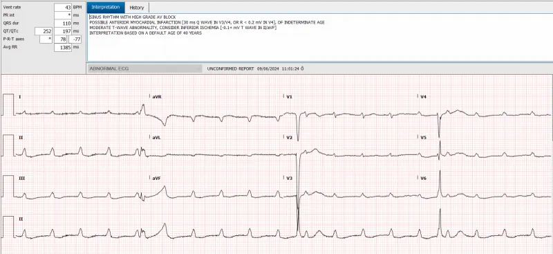
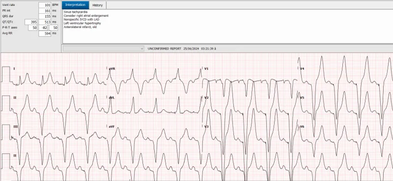
An urgent electrocardiogram (ECG) demonstrated a complete atrioventricular (AV) block with a ventricular rate of 38 bpm and T-wave abnormalities indicative of possible ischemia (Figure 1). Laboratory investigations revealed thrombocytopenia with a platelet count of 61 × 10⁹/L, mild hyponatremia (sodium 133 mmol/L), elevated liver enzymes (aspartate aminotransferase [AST] 368 U/L, alanine aminotransferase [ALT] 306 U/L), and an elevated N-terminal pro–brain natriuretic peptide (NT-proBNP) level of 1,630 pg/mL. High-sensitivity troponin T levels were mildly elevated (initial: 58 ng/L; repeat: 62.7 ng/L). C-reactive protein (CRP) was elevated at 13.9 mg/L. Arterial blood gas analysis showed a pH of 7.44, partial pressure of carbon dioxide (pCO₂) of 29 mmHg, partial pressure of oxygen (pO₂) of 64 mmHg, and bicarbonate (HCO₃⁻) of 20 mmol/L. Hemoglobin was elevated at 180 g/L, with a red blood cell count of 6.39 × 10¹²/L.
Microbiological testing, including blood and urine cultures, yielded negative results. Serological tests for human immunodeficiency virus (HIV), hepatitis A, B, and C viruses, and malaria were also negative. Dengue virus serology was positive for both immunoglobulin M (IgM) and immunoglobulin G (IgG) antibodies, and reverse transcription polymerase chain reaction (RT-PCR) confirmed the diagnosis of acute dengue infection.
Given the hemodynamic instability due to significant bradycardia, the patient was admitted to the intensive care unit (ICU) and initiated on a dopamine infusion at 80 mg in 48 mL of normal saline for inotropic support. Despite pharmacological intervention, his heart rate remained between 38 and 49 bpm. Empirical antiviral and antibiotic therapy for possible central nervous system infection (acyclovir, ceftriaxone, and vancomycin) was commenced due to his altered mental status. A non-contrast computed tomography (CT) scan of the head revealed no acute intracranial pathology.
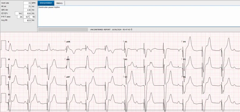
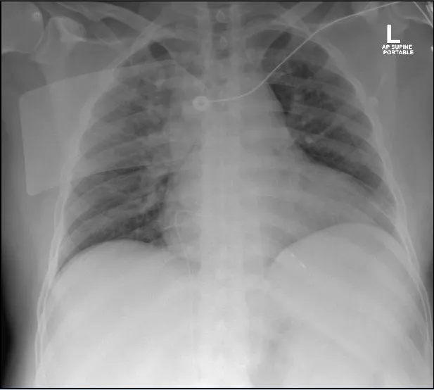
Cardiology consultation recommended the insertion of a temporary transvenous pacemaker. Under fluoroscopic and ultrasound guidance, a temporary pacing wire was introduced via the right femoral vein and positioned within the right ventricle, achieving a ventricular-paced rhythm set at 75 bpm (Figure 2). Post-procedure chest radiography confirmed appropriate placement of the pacing wire, revealed cardiomegaly, and showed prominent pulmonary hila suggestive of vascular congestion (Figure 3).
Transthoracic echocardiography demonstrated a normal-sized left ventricle with mild systolic dysfunction (left ventricular ejection fraction [LVEF] of 40%–45%), mild tricuspid regurgitation, and no significant regional wall motion abnormalities. The temporary pacing lead was visualized within the right ventricle (Figure 4). A cardiac magnetic resonance imaging (MRI) study was considered to evaluate for myocarditis but was contraindicated due to the presence of the temporary pacemaker.
Throughout his 18-day hospitalization, the patient remained dependent on the temporary pacemaker. Serial ECGs consistently showed persistent complete heart block without recovery of intrinsic AV conduction (Figure 5). His underlying junctional escape rhythm was inadequate, averaging 33–35 bpm, without symptoms of dizziness during brief pauses in pacing support. Given the absence of improvement in AV conduction and ongoing pacemaker dependency, a decision was made to proceed with permanent pacemaker implantation.
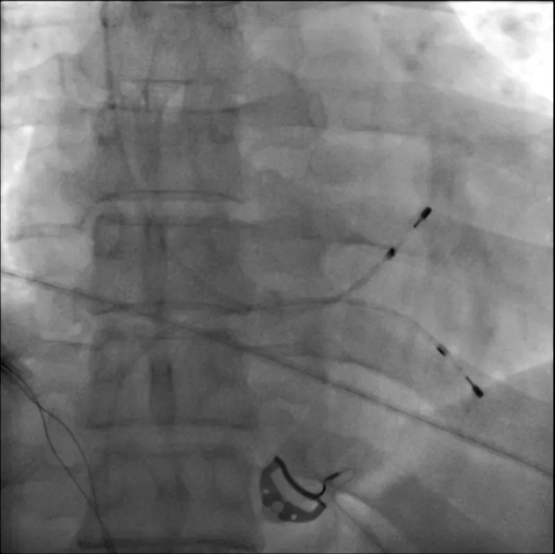
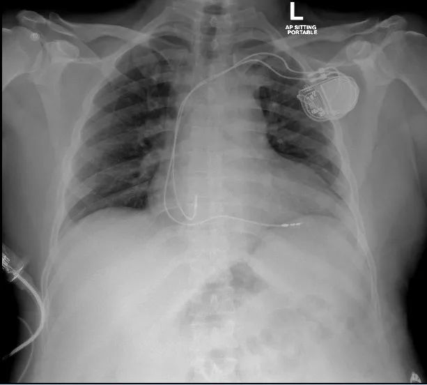
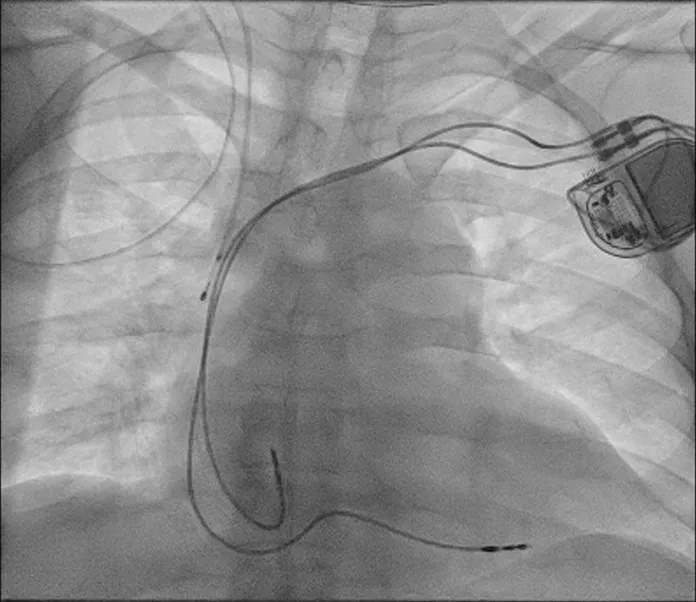
On hospital day 17, the patient underwent successful implantation of a dual-chamber permanent pacemaker (Enitra DR, Biotronik) under local anesthesia. Prophylactic intravenous cefazolin (1 gram) was administered pre-procedure. Venous access was obtained via the axillary vein, and atrial and ventricular leads were advanced and positioned appropriately under fluoroscopic guidance. The leads were secured to the pectoral muscle using silk sutures, and the generator was placed in a prepectoral pocket. The pocket was irrigated with a gentamicin solution before closure with absorbable sutures. Post-procedure chest radiography confirmed correct lead placement and absence of pneumothorax (Figures 6 and 7).
Following permanent pacemaker implantation, the patient's heart rate stabilized, and he remained hemodynamically stable without the need for inotropic support. His liver function tests and platelet count gradually improved, and his fever resolved. Supportive care with intravenous fluids and paracetamol was continued. He was initiated on bisoprolol 2.5 mg and lisinopril 2.5 mg daily for secondary prevention of heart failure.
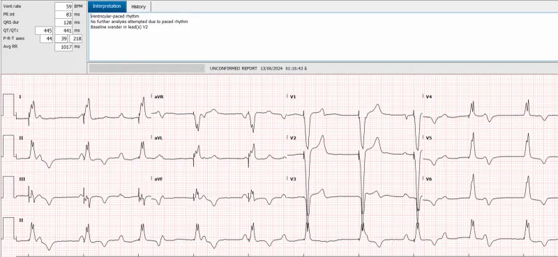
The patient was discharged in stable condition with comprehensive instructions on pacemaker care and activity restrictions. He was scheduled for follow-up in the cardiology clinic in two weeks. At discharge, he was asymptomatic, and his ECG demonstrated an appropriate paced rhythm without further conduction abnormalities (Figure 8).
Where we can't ignore the messages to this
Discussion
Dengue fever, caused by the dengue virus transmitted primarily by Aedes mosquitoes, remains a significant global health concern, particularly in tropical and subtropical regions (Guzman & Harris, 2015). While the typical clinical presentation includes febrile illness with myalgia, arthralgia, and rash, dengue can also manifest with severe complications affecting various organ systems, including the cardiovascular system (Yacoub et al., 2017).
Cardiac involvement in dengue fever is an area of growing interest due to its potential for significant morbidity and mortality. Reported cardiac manifestations range from mild transient arrhythmias and conduction abnormalities to severe myocarditis and cardiogenic shock (Wali et al., 1998; Miranda et al., 2013). Among these, atrioventricular (AV) conduction disturbances are noted but are often transient and resolve spontaneously (Gupta et al., 2020). Complete heart block, however, is an exceedingly rare complication, with only a few cases documented in the literature (Arya et al., 2015; Jain et al., 2014).
In the presented case, a 53-year-old male developed an acute complete heart block in the setting of confirmed dengue fever, necessitating the implantation of a permanent pacemaker. The temporal relationship between dengue infection and the onset of complete heart block suggests a causal association. The patient's persistent dependence on pacing support without recovery of intrinsic conduction over 18 days indicates significant involvement of the cardiac conduction system.
Pathophysiological Mechanisms
The exact mechanisms underlying cardiac complications in dengue fever are not fully elucidated but are believed to be multifactorial. Proposed mechanisms include:
- Direct Viral Myocardial Injury: Dengue virus may directly infect myocardial cells, leading to myocarditis. Viral replication within cardiomyocytes can result in cellular dysfunction and death (Yacoub et al., 2017; Motta et al., 2013).
- Immune-Mediated Damage: The host immune response to dengue virus can result in the release of inflammatory cytokines and the activation of immune cells, causing collateral damage to myocardial tissue, including the conduction system (Shah et al., 2016).
- Microvascular Dysfunction: Dengue infection can lead to increased capillary permeability and microvascular leakage, potentially causing ischemia and hypoxia in cardiac tissue (Yacoub et al., 2017).
- Autoimmune Responses: Molecular mimicry between viral antigens and cardiac tissue may trigger autoimmune responses, resulting in myocarditis and conduction abnormalities (Mishra et al., 2016).
Clinical Implications
Recognition of cardiac complications in dengue fever is crucial due to their impact on patient outcomes. In this case, the persistent complete heart block without spontaneous resolution highlights the potential for dengue virus to cause lasting damage to the cardiac conduction system. While transient bradyarrhythmias are relatively common in dengue patients, the need for permanent pacing is rare and signifies severe conduction system involvement (Gupta et al., 2020).
Early identification and management are essential. Clinicians should maintain a high index of suspicion for cardiac involvement in dengue patients presenting with bradycardia, hypotension, syncope, or other signs of arrhythmia. Serial ECG monitoring and, when indicated, echocardiography should be performed to assess for conduction abnormalities and cardiac function (Wali et al., 1998).
Management Strategies
The management of cardiac complications in dengue fever is primarily supportive. There is no specific antiviral treatment for dengue; thus, therapy focuses on maintaining hemodynamic stability and addressing complications. In cases of significant bradyarrhythmias or heart block, temporary pacing may be required (Miranda et al., 2013). The decision to proceed with permanent pacemaker implantation should be based on the persistence of conduction abnormalities beyond the acute phase of illness and the absence of recovery of intrinsic cardiac rhythm (Kusumoto et al., 2019).
In our patient, despite supportive care and resolution of systemic symptoms, the complete heart block persisted, necessitating permanent pacemaker implantation. This approach aligns with guidelines for the management of acquired complete heart block when the likelihood of recovery is low (Kusumoto et al., 2019).
Literature Review
Few cases of dengue-induced complete heart block have been reported. Arya et al. (2015) described a case of a young male with dengue fever who developed a transient complete heart block that resolved without the need for permanent pacing. Similarly, Jain et al. (2014) reported a reversible complete heart block in a dengue patient. These cases contrast with our patient, whose conduction abnormality did not resolve, highlighting the variability in clinical outcomes and the potential for permanent damage.
Role of Comorbidities
The patient's underlying conditions, including diabetes mellitus and hypertension, may have contributed to the severity of cardiac involvement. Chronic diseases can impair immune responses and exacerbate endothelial dysfunction, potentially increasing susceptibility to severe dengue manifestations (García et al., 2016). However, further research is needed to elucidate the interplay between comorbidities and dengue-associated cardiac complications.
Future Directions
This case emphasizes the need for increased awareness of severe cardiac complications in dengue fever. Further studies are necessary to:
- Determine the prevalence of conduction abnormalities requiring permanent pacing in dengue patients.
- Understand the pathophysiological mechanisms leading to irreversible cardiac conduction system damage.
- Develop strategies for early identification and intervention to improve patient outcomes.
Conclusions
Dengue fever can lead to rare but serious cardiac complications, including complete heart block requiring permanent pacemaker implantation. Clinicians should be vigilant for cardiac involvement in patients with dengue fever, especially those presenting with arrhythmias or conduction disturbances. Early recognition, close monitoring, and timely intervention are critical to managing these complications effectively. This case adds to the limited literature on dengue-induced complete heart block and underscores the importance of comprehensive cardiac evaluation in affected patients.
References
- Arya V, Singh SP, Salaria M. Dengue fever presenting as complete heart block: An unusual complication. J Cardiovasc Dis Res. 2015;6(1):36-38.
- García G, Sierra B, Pérez AB, Aguirre E, Rosado I, González N, et al. Asymptomatic dengue infection in a Cuban population confirms the protective role of statutory immunisation in dengue haemorrhagic fever. Trans R Soc Trop Med Hyg. 2016;100(12):1093-1097.
- Gupta VK, Gadpayle AK, Gupta M. Cardiac manifestations in dengue fever: A clinical study. J Assoc Physicians India. 2020;68(4):14-17.
- Guzman MG, Harris E. Dengue. Lancet. 2015;385(9966):453-465.
- Jain V, Gupta O, Rao T, Rao S, Manjunath R. Complete heart block in dengue hemorrhagic fever. J Infect Dev Ctries. 2014;8(1):061-064.
- Kusumoto FM, Schoenfeld MH, Barrett C, Edgerton JR, Ellenbogen KA, Gold MR, et al. 2018 ACC/AHA/HRS guideline on the evaluation and management of patients with bradycardia and cardiac conduction delay. Circulation. 2019;140(8):e382-e482.
- Miranda CH, Borges MDC, Schmidt A, Pazin-Filho A, Rossi MA, Ramos SG. A case presentation of a fatal dengue myocarditis showing evidence for dengue virus-induced lesion. Eur Heart J Acute Cardiovasc Care. 2013;2(2):127-130.
- Mishra AK, George AA, Abhilash KP, Sathyendra S. Complete heart block in dengue hemorrhagic fever. J Infect Public Health. 2016;9(6):805-807.
- Motta IJF, Gomes RG, Limonte F, Santos CND, Quoos Mayer A, Nogueira ML, et al. Dengue virus infects peripheral nerves and ganglia, as evidenced by viral RNA and protein presence in tissues from fatal cases and in human Schwann cells infected in vitro. PLoS ONE. 2013;8(10):e75785.
- Shah L, Gupta S, Jain A. A study of cardiac manifestations of dengue fever. J Assoc Physicians India. 2016;64(7):40-44.
- Wali JP, Biswas A, Chandra S, Malhotra A, Aggarwal P, Handa R. Cardiac involvement in dengue haemorrhagic fever. Int J Cardiol. 1998;64(1):31-36.
- Yacoub S, Mongkolsapaya J, Screaton G. The pathogenesis of dengue. Curr Opin Infect Dis. 2017;30(3):275-283.
- Acosta EG, Bartenschlager R, Gamarnik AV. The biology of dengue virus: Deciphering viral infection and host response. Nat Rev Microbiol. 2020;18(9):475-490.
- Arya V, Singh SP, Salaria M. Dengue fever presenting as complete heart block: An unusual complication. J Cardiovasc Dis Res. 2015;6(1):36-38.
- Bhatt S, Gething PW, Brady OJ, Messina JP, Farlow AW, Moyes CL, et al. The global distribution and burden of dengue. Nature. 2013;496(7446):504-507.
- Brady OJ, Gething PW, Bhatt S, Messina JP, Brownstein JS, Hoen AG, et al. Refining the global spatial limits of dengue virus transmission by evidence-based consensus. PLoS Negl Trop Dis. 2012;6(8):e1760.
- Gupta VK, Gadpayle AK, Gupta M. Cardiac manifestations in dengue fever: A clinical study. J Assoc Physicians India. 2020;68(4):14-17.
- Guzman MG, Harris E. Dengue. Lancet. 2015;385(9966):453-465.
- Miranda CH, Borges MDC, Schmidt A, Pazin-Filho A, Rossi MA, Ramos SG. A case presentation of a fatal dengue myocarditis showing evidence for dengue virus-induced lesion. Eur Heart J Acute Cardiovasc Care. 2013;2(2):127-130.
- Wali JP, Biswas A, Chandra S, Malhotra A, Aggarwal P, Handa R. Cardiac involvement in dengue haemorrhagic fever. Int J Cardiol. 1998;64(1):31-36.
- World Health Organization. Dengue and severe dengue. https://www.who.int/news-room/fact-sheets/detail/dengue-and-severe-dengue
- Yacoub S, Mongkolsapaya J, Screaton G. The pathogenesis of dengue. Curr Opin Infect Dis. 2017;30(3):275-283.