
Sonographic Characterization of Adenomyosis Using the Morphological Uterus Sonographic Assessment (MUSA) Criteria and Its Association With Clinical Presentation and Disease Severity: A Retrospective Cohort Study
Authors

Published
October 17, 2026
Paper ID
PAP-000319
Status
Published
DOI
10.65712/tmj.2093276
Advance Services
Abstract
Background and Objective: Transvaginal ultrasonography utilizing the Morphological Uterus Sonographic Assessment (MUSA) criteria is the standard for diagnosing adenomyosis, yet the precise relationship between specific sonographic phenotypes and clinical severity remains under-investigated. This study aimed to evaluate the clinical relevance of ultrasound-based characterization of adenomyosis using MUSA features and to examine its associations with specific gynecological symptoms and markers of disease severity.
Methods: This single-center, retrospective cohort study included 100 premenopausal symptomatic women diagnosed with adenomyosis via transvaginal ultrasound at a tertiary referral center. Sonographic evaluation documented the presence of direct and indirect MUSA features, adenomyosis subtype (diffuse, focal, or @@mixed), uterine morphometry, and total MUSA feature count. Bivariate and multivariable analyses were utilized to quantify associations between individual sonographic features, presenting symptoms (e.g., heavy menstrual bleeding, dysmenorrhea), and secondary clinical severity markers (e.g., anemia, intravenous iron therapy, and emergency samrt heart.
Results: The clinical profile of the cohort was dominated by abnormal uterine bleeding, with 71% of patients reporting heavy menstrual bleeding (HMB), which drove a substantial hematologic and healthcare burden. Sonographically, patients exhibited a high lesion burden with a mean of 6.5 MUSA features per patient. Analysis of specific feature-symptom pairings revealed that fan-shaped shadowing and an irregular junctional zone (JZ) were significantly associated with HMB, while the presence of a coexisting endometrioma correlated with intermenstrual bleeding. An inverse association was found between JZ irregularity and deep dyspareunia. HMB emerged as the principal driver of disease severity, strongly correlating with anemia (p < 0.001), the need for intravenous iron (p < 0.001), and emergency visits for bleeding (p < 0.001). Multivariable analysis confirmed that both HMB and the diffuse adenomyosis subtype are independent predictors of anemia. Quantitative measures, including uterine volume and total MUSA feature count, were not significantly associated with levonorgestrel-releasing intrauterine system (LNG-IUS) failure.
Conclusion: Specific sonographic MUSA features—particularly fan-shaped shadowing and junctional zone irregularity—are valuable diagnostic indicators of heavy menstrual bleeding and overall clinical severity in patients with adenomyosis. Recognizing these specific ultrasound phenotypes tracks more closely with abnormal bleeding than pain, allowing clinicians to better stratify patients at higher risk for severe hematologic complications.
Introduction
Adenomyosis is a benign gynecological condition characterized by the abnormal presence of endometrial glands and stroma within the myometrium.1 Historically, the disease was primarily diagnosed retrospectively through the histological examination of hysterectomy specimens, which biased the epidemiological understanding of the condition towards older, parous women.1,2 However, recent advancements in imaging have fundamentally shifted this paradigm, establishing transvaginal ultrasonography (TVS) as a highly accurate, non-invasive, and accessible first-line modality for diagnosis.1,3 To standardize the reporting of these sonographic findings, the Morphological Uterus Sonographic Assessment (MUSA) group introduced consensus definitions for ultrasound features typical of adenomyosis.4 These key features include an inhomogeneous myometrium, asymmetrical thickening, myometrial cysts, hyperechogenic islands, fan-shaped shadowing, and an irregular or interrupted junctional zone (JZ),4 which have recently been further categorized into direct and indirect signs of the disease.5
Clinically, adenomyosis presents with a heterogeneous and often debilitating spectrum of symptoms, most notably abnormal uterine bleeding (AUB)—especially heavy menstrual bleeding (HMB)—and severe pelvic pain, including dysmenorrhea and dyspareunia.1 Despite the robust standardization of MUSA criteria, the precise relationship between specific sonographic phenotypes and clinical severity remains a subject of ongoing investigation.5 Previous studies have demonstrated significant correlations between the total number of sonographic features and symptom severity.2,6 For instance, Naftalin et al.6 established a positive correlation between the number of TVS features and the severity of menstrual pain. Similarly, Pinzauti et al.2 found that the number of adenomyotic features correlated with both heavy menstrual bleeding and dysmenorrhea in younger populations. Conversely, other research suggests that different patterns of the disease, such as diffuse adenomyosis, correlate more strongly with older age and heavier menstrual bleeding.7
Given the variability in clinical presentations, there is a distinct need to further elucidate how specific MUSA features correlate with presenting symptoms and broader markers of disease burden. Therefore, this retrospective cohort study was designed to evaluate the clinical relevance of ultrasound-based characterization of adenomyosis using the MUSA consensus criteria, and to examine its association with specific gynecological symptoms and markers of disease severity in premenopausal women.
Materials and Methods
Study Design and Setting
This was a single-centre, retrospective cohort study conducted at Corniche Hospital, a tertiary maternal and neonatal referral centre in Abu Dhabi, United Arab Emirates. Consecutive premenopausal women who underwent transvaginal ultrasonography and received a sonographic diagnosis of adenomyosis between January 2022 and December 2025 were eligible. The study was designed to evaluate the clinical relevance of ultrasound-based characterisation of adenomyosis according to the Morphological Uterus Sonographic Assessment (MUSA) consensus and to examine its association with the presence of gynaecological symptoms and with markers of disease severity. The final analytic sample comprised 100 women.
Participants and Eligibility Criteria
Eligible participants were women aged 18 to 50 years who were managed at the institution and whose transvaginal ultrasound examinations were performed within its imaging facilities. A diagnosis of adenomyosis established by trained sonographers on transvaginal ultrasound according to MUSA features was required, and each woman had to present with at least one gynaecological symptom, namely dysmenorrhoea, heavy menstrual bleeding, chronic pelvic pain, or dyspareunia. Women were excluded if they were postmenopausal (amenorrhoea exceeding 12 months); had undergone hysterectomy or any adenomyosis-directed intervention before the baseline scan; had a diagnosis based solely on magnetic resonance imaging without transvaginal ultrasound confirmation; or had incomplete sonographic or clinical documentation. Further exclusions comprised coexisting uterine fibroids, congenital uterine anomalies, suspected gynaecological malignancy, previously diagnosed severe endometriosis, chronic pain of non-gynaecological origin, and the use of hormonal therapy before baseline symptom and ultrasound assessment.
Data Sources and Extraction
Clinical, demographic, and imaging data were extracted from the hospital electronic health record (Cerner) by the Health Information Management department using International Classification of Diseases codes, clinical encounter records, structured clinical documentation, and stored ultrasound images and reports. Extracted data were compiled into a single password-protected spreadsheet to which access was restricted to the research team.
Sonographic Assessment and MUSA Classification
Transvaginal ultrasound examinations were performed and reported by trained sonographers. Adenomyosis was characterised using the direct and indirect MUSA features: myometrial cysts or lacunae, hyperechogenic islands, echogenic subendometrial lines and buds, a heterogeneous (inhomogeneous) myometrium, asymmetrical myometrial thickening, fan-shaped shadowing, a globular uterine configuration, junction-zone morphology, and translesional vascularity; the presence of a coexisting endometrioma was also recorded. On the basis of lesion distribution, each uterus was classified as diffuse, focal, or mixed adenomyosis. Uterine dimensions (longitudinal, anteroposterior, and transverse diameters), uterine volume, and endometrial thickness were measured, and the overall sonographic burden was summarised as a MUSA feature count.
Study Variables and Outcome Definitions
The recorded variables spanned four domains. Demographic and anthropometric variables comprised age, height, weight, and body mass index. Obstetric and menstrual variables comprised parity, previous caesarean section, previous miscarriage, previous surgical management of miscarriage, previous oral contraceptive use, menstrual-cycle regularity, and menstrual duration. Clinical symptoms comprised heavy menstrual bleeding, intermenstrual bleeding, dysmenorrhoea, deep dyspareunia, pelvic pain, dyschezia, and urinary symptoms. Sonographic variables comprised the individual MUSA features, the adenomyosis subtype, uterine morphometry, and the MUSA feature count. The primary outcome was the association between individual MUSA features and the presence of each gynaecological symptom. Secondary outcomes were the demographic, obstetric, and clinical correlates of the sonographic phenotype; the association of sonographic features with failure of the levonorgestrel-releasing intrauterine system (LNG-IUS), defined as device removal or expulsion within 12 months of insertion; and indicators of disease severity, namely anaemia (haemoglobin 10 g/dL or below), intravenous iron therapy, blood transfusion, and emergency attendance or admission for pelvic pain or for abnormal uterine bleeding.
Data Handling and Quality Control
Before analysis, the dataset underwent structured cleaning. Free-text and mixed-format entries were standardised: ages recorded as text were parsed to integers, and category labels were harmonised for case and spelling. Haemoglobin values recorded in mixed units and as multiple readings per patient were harmonised to grams per decilitre, and the lowest (nadir) value per patient was retained to reflect the most severe anaemia. One biologically implausible anteroposterior uterine diameter and a small number of malformed measurement entries were treated as missing. The MUSA feature count was derived as the number of sonographic features present per patient. Two indirect features, a heterogeneous myometrium and hyperechogenic islands, were present in every participant and therefore carried no discriminative information; these were retained in the descriptive summary but excluded from the association analyses. Variables with missing values were handled by available-case analysis, and the denominator is reported wherever it differed from the full sample.
Statistical Analysis
Continuous variables were summarised as mean (standard deviation) and categorical variables as frequency (percentage). Associations between categorical variables were examined using the chi-square or Fisher exact test, and continuous variables were compared between groups using the independent-samples t-test or the Mann–Whitney U test, as appropriate to the distribution. For the primary outcome, the association between each MUSA feature and each symptom was quantified by the odds ratio with 95% confidence interval obtained from two-by-two contingency analysis with the Haldane–Anscombe correction, and statistical significance was assessed by the Fisher exact test. Associations between continuous predictors and binary outcomes were estimated by univariable logistic regression, and correlations between continuous variables by the Spearman rank coefficient. To identify independent predictors of anaemia, a multivariable logistic regression model was fitted with heavy menstrual bleeding, uterine volume, the MUSA feature count, and diffuse subtype as covariates. All tests were two-tailed, and a p-value below 0.05 was considered statistically significant. Analyses were performed in Python using the pandas, SciPy, and statsmodels libraries.
Ethical Considerations
The study was conducted in accordance with the principles of the Declaration of Helsinki. The protocol was reviewed and approved by the institutional research ethics committee (approval number: [to be inserted]), and the requirement for individual informed consent was waived in view of the retrospective design and the use of de-identified data. All records were anonymised and stored securely, with access restricted to the research team.
Results
Cohort and Clinical Profile
The cohort comprised 100 premenopausal women in the later reproductive years, the majority of whom were parous and fell within the overweight or obese range of body mass index (Table 1). Clinical presentation was dominated by abnormal uterine bleeding rather than by pain: heavy menstrual bleeding was by far the most frequent complaint, whereas deep dyspareunia, dyschezia, and urinary symptoms were uncommon. The haematologic and healthcare burden was substantial, with a sizeable minority of women anaemic and a meaningful proportion requiring intravenous iron or unscheduled hospital attendance for bleeding, underscoring the clinical impact of the disease in this population.
| Characteristic | Value |
|---|---|
| Demographic and anthropometric | |
| Age, years | 43.7 (4.5) |
| Height, cm | 160.1 (5.3) |
| Weight, kg | 77.1 (14.5) |
| Body mass index, kg/m² | 30.3 (6.2) |
| Obstetric and menstrual history | |
| Parity, mean (SD) | 4.3 (1.9) |
| Previous cesarean section | 50 (50.0) |
| Previous miscarriage | 26 (26.0) |
| Surgical management of miscarriage | 15 (15.0) |
| Previous oral contraceptive use | 15 (15.0) |
| Regular menstrual cycle | 59 (59.0) |
| Menstruation length > 8 days | 38 (38.0) |
| Gynaecological symptoms | |
| Heavy menstrual bleeding | 71 (71.0) |
| Intermenstrual bleeding | 26 (26.0) |
| Dysmenorrhea | 43 (43.0) |
| Deep dyspareunia | 8 (8.0) |
| Pelvic pain | 25 (25.0) |
| Dyschezia | 1 (1.0) |
| Urinary symptoms | 10 (10.0) |
| Haematologic and healthcare burden | |
| Haemoglobin ≤ 10 g/dL | 32 (33.0) |
| Haemoglobin (nadir), g/dL | 10.8 (1.8) |
| Intravenous iron therapy | 24 (24.7) |
| Blood transfusion | 3 (3.1) |
| Emergency visit/admission for pelvic pain | 10 (10.1) |
| Emergency visit/admission for abnormal uterine bleeding | 27 (27.0) |
Note. Continuous variables are reported as mean (SD) and categorical variables as n (%), to one decimal place; percentages appear in parentheses and are based on non-missing observations. Body mass index was available for 96 patients. The haemoglobin ≤ 10 g/dL indicator was available for 97 patients; the haemoglobin nadir (lowest recorded value, converted to g/dL) was available for 84 patients. SD = standard deviation.
Sonographic Phenotype
On ultrasound, the cohort was characterised by a high overall lesion burden (Table 2). The indirect features of a heterogeneous myometrium and hyperechogenic islands were universal, whereas the direct features of myometrial cysts and subendometrial lines and buds were present in only a minority, indicating that the diagnosis rested predominantly on indirect signs. Diffuse disease was the most frequent subtype, an abnormal junction zone was present in most uteri, and lesional translesional vascularity was identified in approximately half. Uterine dimensions and volume were consistent with a generally enlarged, globular uterus, in keeping with established disease.
| Sonographic parameter | Value |
|---|---|
| Qualitative MUSA features, n (%) | |
| Inhomogeneous/heterogeneous myometrium | 100 (100.0) |
| Myometrial cysts/lacunae | 38 (38.0) |
| Hyperechogenic islands | 100 (100.0) |
| Echogenic subendometrial lines and buds | 35 (35.0) |
| Asymmetrical myometrial thickening | 90 (90.0) |
| Fan-shaped shadowing | 82 (82.0) |
| Globular uterus | 78 (78.0) |
| Endometrioma | 5 (5.0) |
| Junction zone, n (%) | |
| Regular | 18 (18.0) |
| Irregular | 36 (36.0) |
| Interrupted | 24 (24.0) |
| Not visible | 22 (22.0) |
| Translesional vascularity, n (%) | |
| Uniform | 33 (33.0) |
| Non-uniform | 28 (28.0) |
| Interlesional | 11 (11.0) |
| Both circular and interlesional | 10 (10.0) |
| Not evaluated | 18 (18.0) |
| Adenomyosis subtype, n (%) | |
| Diffuse | 52 (52.0) |
| Focal | 15 (15.0) |
| Mixed | 33 (33.0) |
| MUSA feature burden | |
| Number of MUSA features per patient, mean (SD) | 6.5 (1.4) |
| Quantitative measurements, mean (SD) | |
| Longitudinal uterine diameter, mm | 82.5 (12.8) |
| Anteroposterior uterine diameter, mm | 53.2 (9.0) |
| Transverse uterine diameter, mm | 60.9 (10.6) |
| Uterine volume, cm³ | 149.7 (72.0) |
| Endometrial thickness, mm | 9.8 (5.7) |
Note. MUSA = Morphological Uterus Sonographic Assessment. Categorical features are reported as n (%) and quantitative measurements as mean (SD), to one decimal place; percentages appear in parentheses. The MUSA feature count is the number of sonographic MUSA features present per patient (range 0–9): inhomogeneous myometrium, hyperechogenic islands, myometrial cysts/lacunae, echogenic subendometrial lines and buds, asymmetrical thickening, fan-shaped shadowing, globular uterus, junction-zone abnormality, and lesional translesional vascularity. One implausible anteroposterior diameter value (883.5 mm) was excluded (n = 99); endometrial thickness was available for 99 patients.
Feature–Symptom Associations (Primary Outcome)
At the level of individual feature–symptom pairings, most associations did not reach statistical significance, and no single MUSA feature tracked uniformly across the symptom spectrum (Figure 1). The significant signals were few but clinically coherent and converged on the bleeding phenotype: fan-shaped shadowing and an irregular junction zone were each positively associated with heavy menstrual bleeding, while a coexisting endometrioma was associated with intermenstrual bleeding. An inverse association was observed between junction-zone irregularity and deep dyspareunia. Associations involving dyschezia could not be meaningfully evaluated because the symptom was reported by a single patient. Taken together, the pattern suggests that, in this cohort, sonographic features were more closely linked to abnormal bleeding than to the pain-predominant presentations.
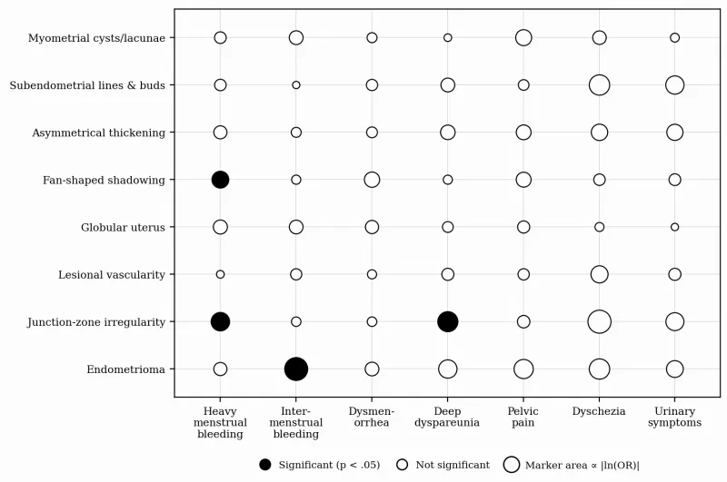
Note. Each marker represents the bivariate association between a MUSA sonographic feature (rows) and a gynaecological symptom (columns), quantified by the odds ratio from a 2 × 2 analysis (Haldane–Anscombe correction; Fisher exact p). Marker area is proportional to the strength of association, |ln(odds ratio)|; filled markers denote statistically significant associations (p < 0.05) and open markers non-significant associations. Inhomogeneous myometrium and hyperechogenic islands were present in all patients and are therefore not shown. The dyschezia column reflects a single affected patient and is statistically uninformative.
Symptom Burden Across Adenomyosis Subtypes
When symptoms were stratified by subtype, the bleeding-predominant pattern was most evident in diffuse and mixed disease and least so in focal disease (Figure 2). Focal disease, by contrast, was comparatively more associated with pelvic pain. Other symptoms were distributed more evenly across the three subtypes, and the small number of women with focal disease warrants caution in interpreting subtype-specific estimates.
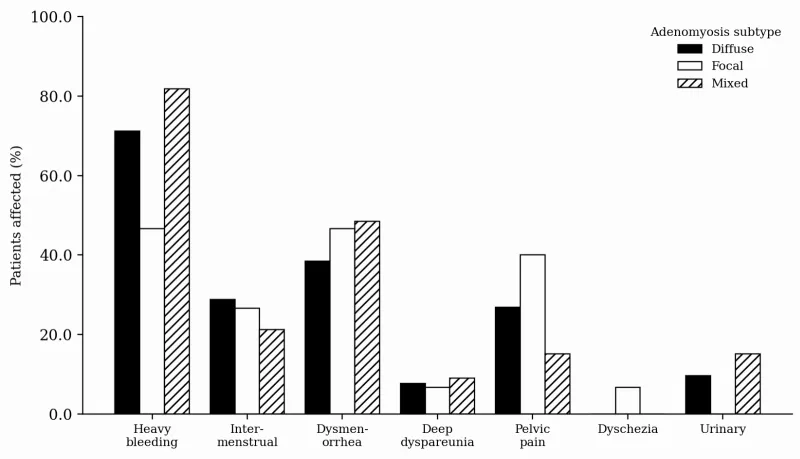
Note. Bars show the percentage of patients reporting each symptom within the diffuse (n = 52), focal (n = 15), and mixed (n = 33) adenomyosis subtypes; subtypes are distinguished by fill pattern.
Secondary Clinical Associations
Among the severity outcomes, heavy menstrual bleeding emerged as the dominant clinical correlate, showing strong and consistent associations with anaemia, the need for intravenous iron, and emergency attendance for abnormal uterine bleeding (Table 3). In contrast, the quantitative sonographic measures, namely uterine volume and the MUSA feature count, and the adenomyosis subtype were not significantly associated with LNG-IUS failure or with the obstetric and demographic factors examined, and the corresponding correlations were weak. Estimates for blood transfusion and for device failure rested on very few events and should therefore be regarded as imprecise.
| Predictor | OR (95% CI) | p |
|---|---|---|
| Anaemia (haemoglobin ≤ 10 g/dL) | ||
| Heavy menstrual bleeding | 5.7 (1.7–19.0) | < .001 |
| Uterine volume (per 50 cm³) | 1.2 (0.9–1.6) | .17 |
| MUSA feature count (per feature) | 1.0 (0.7–1.4) | .98 |
| Diffuse subtype (vs non-diffuse) | 2.3 (1.0–5.5) | .06 |
| Intravenous iron therapy | ||
| Heavy menstrual bleeding | 9.8 (1.8–54.6) | < .001 |
| Uterine volume (per 50 cm³) | 1.0 (0.8–1.4) | .78 |
| Diffuse subtype (vs non-diffuse) | 2.8 (1.1–7.4) | .04 |
| Blood transfusion (n = 3 events; interpret with caution) | ||
| Heavy menstrual bleeding | 3.2 (0.2–63.0) | .55 |
| Anaemia (Hb ≤ 10 g/dL) | 3.6 (0.5–28.9) | .24 |
| LNG-IUS failure within 12 months (n = 8 events; interpret with caution) | ||
| Diffuse subtype (vs non-diffuse) | 1.5 (0.4–6.1) | .72 |
| Uterine volume (per 50 cm³) | 1.1 (0.7–1.7) | .74 |
| MUSA feature count (per feature) | 1.0 (0.6–1.7) | .96 |
| Globular uterus | 0.8 (0.2–3.5) | 1.00 |
| Emergency visit/admission for abnormal uterine bleeding | ||
| Heavy menstrual bleeding | 6.0 (1.5–24.0) | < .001 |
| Uterine volume (per 50 cm³) | 1.1 (0.8–1.5) | .40 |
| Obstetric and demographic factors | ||
| Previous cesarean section: uterine volume (per 50 cm³) | 0.8 (0.6–1.1) | .20 |
| Previous miscarriage: diffuse subtype | 1.1 (0.5–2.7) | 1.00 |
| Globular uterus: body mass index (per kg/m²) | 1.0 (0.9–1.0) | .24 |
| Age × MUSA feature count | ρ = 0.2 | .02 |
| Parity × uterine volume | ρ = 0.0 | .87 |
| Body mass index × uterine volume | ρ = 0.0 | .83 |
Note. Effect sizes are odds ratios (OR) with 95% confidence intervals (CI) from bivariate logistic regression (continuous predictors) or 2 × 2 analysis with Fisher exact test (binary predictors); ρ denotes the Spearman rank correlation for continuous–continuous associations. Values are reported to one decimal place; all tests were two-tailed, with p < 0.05 considered statistically significant. Outcomes with few events (blood transfusion, n = 3; LNG-IUS failure, n = 8) yield unstable estimates and should be interpreted with caution. LNG-IUS = levonorgestrel-releasing intrauterine system.
Independent Predictors of Anaemia
In the multivariable model, the bleeding phenotype retained its prominence (Figure 3). Heavy menstrual bleeding was independently associated with anaemia, as was the diffuse subtype, whereas the overall feature count and uterine volume contributed little once bleeding and subtype were taken into account. The directional consistency between the bivariate and adjusted analyses strengthens the inference that the bleeding-predominant, diffuse phenotype carries the greatest haematologic risk, although the modest number of events limits the precision of the adjusted estimates.
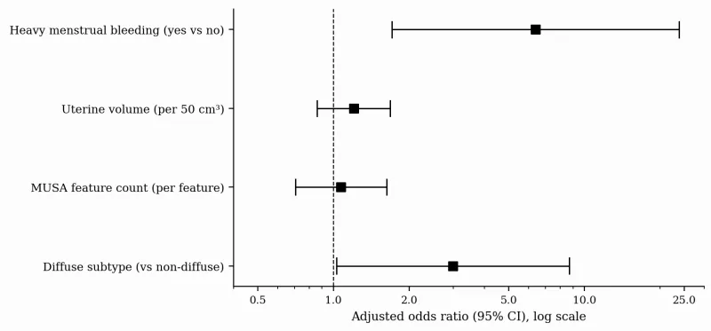
Note. Squares represent adjusted odds ratios and horizontal lines the 95% confidence intervals from a multivariable logistic regression model of anaemia (haemoglobin ≤ 10 g/dL; n = 97, 32 events); the dashed vertical line marks the null value (OR = 1.0). With four predictors and 32 events, the model is modestly powered (approximately eight events per variable) and the estimates should be regarded as exploratory.
Discussion
Our study demonstrates that in a cohort of premenopausal women, specific sonographic features of adenomyosis track more closely with abnormal bleeding phenotypes than with pain-predominant presentations. At the level of individual features, fan-shaped shadowing and an irregular junctional zone (JZ) were significantly associated with heavy menstrual bleeding (HMB), while the presence of a coexisting endometrioma correlated with intermenstrual bleeding. Intriguingly, we observed an inverse association between JZ irregularity and deep dyspareunia. Furthermore, HMB emerged as the principal driver of clinical disease severity, demonstrating strong and consistent associations with anemia, the requirement for intravenous iron therapy, and emergency hospital visits for abnormal uterine bleeding. However, quantitative sonographic measures, including the total MUSA feature count and uterine volume, were not significantly associated with levonorgestrel-releasing intrauterine system (LNG-IUS) failure or other severity outcomes.
These findings provide an important contrast to some existing literature that strongly links the overall sonographic burden of adenomyosis to pain severity. For example, Naftalin et al.6 demonstrated a direct, statistically significant positive correlation between the number of ultrasound features of adenomyosis and the severity of menstrual pain. In our cohort, no single MUSA feature tracked uniformly across the symptom spectrum, and the significant signals converged primarily on bleeding. Our results align more closely with the findings of Exacoustos et al.,7 who reported that diffuse sonographic patterns of adenomyosis were significantly associated with heavier menstrual bleeding. The discrepancies between studies may reflect differences in cohort demographics, overlapping pathologies such as fibroids or endometriosis, or varying degrees of disease progression.1,7
The specific association of an irregular JZ and fan-shaped shadowing with HMB is pathophysiologically plausible. The JZ plays a critical role in coordinating uterine contractility.8,9 Thickening, disruption, or irregularity of the JZ architecture—increasingly recognized as a hallmark of adenomyosis—can alter the coordinated peristaltic activity of the inner myometrium, thereby impairing the mechanisms required to control menstrual blood loss.8,9 Harmsen et al.5 highlighted the importance of JZ assessment, classifying irregular and interrupted JZ as key indirect sonographic signs of the disease.
The identification of distinct feature-symptom pairings carries important clinical implications. Since HMB was the dominant driver of hematologic and healthcare burden in our study, leading to anemia and unscheduled hospital admissions, recognizing specific sonographic markers like fan-shaped shadowing or JZ irregularity could help clinicians stratify patients at higher risk for severe bleeding complications. This risk stratification may prompt earlier and more targeted medical interventions. The lack of correlation between uterine volume or feature count and LNG-IUS failure suggests that this therapeutic option remains viable regardless of the extensive sonographic appearance of the disease.
Strengths of this study include the standardized characterization of adenomyosis using the updated MUSA criteria by trained sonographers, as well as the comprehensive evaluation of secondary disease severity markers. Limitations include the retrospective, single-center design, which may introduce selection bias, and the reliance on a symptom profile that may be influenced by unmeasured confounding factors.
Conclusions
Findings suggest that individual MUSA features—particularly fan-shaped shadowing and junctional zone irregularity—are valuable indicators of abnormal uterine bleeding and clinical severity in women with adenomyosis. Further prospective, multi-center studies are warranted to validate these specific feature-symptom associations and to determine their utility in guiding therapeutic decision-making.
References
- Chapron C, Vannuccini S, Santulli P, Abrão MS, Carmona F, Fraser IS, et al. Diagnosing adenomyosis: an integrated clinical and imaging approach. Hum Reprod Update. 2020;26(3):392-411.
- Pinzauti S, Lazzeri L, Tosti C, Centini G, Orlandini C, Luisi S, et al. Transvaginal sonographic features of diffuse adenomyosis in 18-30-year-old nulligravid women without endometriosis: association with symptoms. Ultrasound Obstet Gynecol. 2015;46(6):730-6.
- Andres MP, Borrelli GM, Ribeiro J, Baracat EC, Abrão MS, Kho RM. Transvaginal ultrasound for the diagnosis of adenomyosis: systematic review and meta-analysis. J Minim Invasive Gynecol. 2018;25(2):257-64.
- Van den Bosch T, Dueholm M, Leone FPG, Valentin L, Rasmussen CK, Votino A, et al. Terms, definitions and measurements to describe sonographic features of myometrium and uterine masses: a consensus opinion from the Morphological Uterus Sonographic Assessment (MUSA) group. Ultrasound Obstet Gynecol. 2015;46(3):284-98.
- Harmsen MJ, Van den Bosch T, de Leeuw RA, Dueholm M, Exacoustos C, Valentin L, et al. Consensus on revised definitions of Morphological Uterus Sonographic Assessment (MUSA) features of adenomyosis: results of a modified Delphi procedure. Ultrasound Obstet Gynecol. 2022;60(1):118-31.
- Naftalin J, Hoo W, Nunes N, Holland T, Mavrelos D, Jurkovic D. Association between ultrasound features of adenomyosis and severity of menstrual pain. Ultrasound Obstet Gynecol. 2016;47(6):779-83.
- Exacoustos C, Morosetti G, Conway F, Camilli S, Martire FG, Lazzeri L, et al. New sonographic classification of adenomyosis: do type and degree of adenomyosis correlate to severity of symptoms? J Minim Invasive Gynecol. 2020;27(6):1308-15.
- Exacoustos C, Brienza L, Di Giovanni A, Szabolcs B, Romanini ME, Zupi E, et al. Adenomyosis: three-dimensional sonographic findings of the junctional zone and correlation with histology. Ultrasound Obstet Gynecol. 2011;37(4):471-9.
- Rasmussen CK, Hansen ES, Dueholm M. Two- and three-dimensional ultrasonographic features related to histopathology of the uterine endometrial-myometrial junctional zone. Acta Obstet Gynecol Scand. 2019;98(2):205-14.